About vistaEKG:
 EKG has been reinvented for the very first time since 1903, 3 big differences between conventional EKG and vistaEKG: 1) qualitative 2) quantitative 3) positioning. Quantitative and qualitative diagnostics are invented for the first time. Here come 4 key initiatives: a) linear waveform is segmented in different colors and the automatic detection, identification, separation, color marking are implemented by artificial intelligence according to the potential of the autonomic conduction system. b) Quantitative data of EKG parameters, including invasive EKG parameters, are navigated and mapped by AI automatically; the accuracy is comparable to invasive EKG monitoring. c) Real time navigation automatically reading system for abnormal morphological wave. Various symbols and arrows indicate a certain heartbeat wave and an abnormal local position. d) Suggestion of clinical experts.
Theory and technology:
EKG signals of the autonomic conduction system are hidden in the P-wave and various electrical signals of membrane potential are hidden in the T-wave, but they cannot be displayed or extracted. vistaEKG achieves the technologies of all those 4 above through the combination of X-Y-Z axis tracking mode, electromagnetic and impedance, frequency and interval, Multiple frequencies and multiple thresholds combination etc. Detailed to each heartbeat, and independent special signal processing technology is applied.

Clinical and Application:
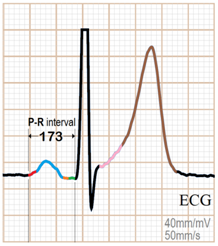
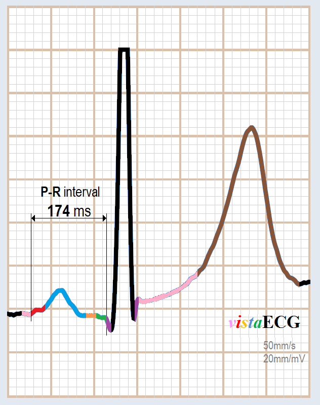
A) Anatomical local visualization of EKG waveforms shows in the P-wave of conventional EKG:

SN→ atrium(red)
→ Atrioventricular node (blue)
→bundle of His(yellow)
→ bundle branch(green)
→ Purkinje's initial point(purple)
→ Purkinje’s terminal point(purple)
→ST segment(pink)
→ T segment(brown)
B) RBBB shows in conventional EKG as CAD+RBBB shows in vistaEKG.

C) Abnormal signal reading system with navigation automatically.

D) Meaning of navigation symbol.

About vistaEKG:
 EKG has been reinvented for the very first time since 1903, 3 big differences between conventional EKG and vistaEKG: 1) qualitative 2) quantitative 3) positioning. Quantitative and qualitative diagnostics are invented for the first time. Here come 4 key initiatives: a) linear waveform is segmented in different colors and the automatic detection, identification, separation, color marking are implemented by artificial intelligence according to the potential of the autonomic conduction system. b) Quantitative data of EKG parameters, including invasive EKG parameters, are navigated and mapped by AI automatically; the accuracy is comparable to invasive EKG monitoring. c) Real time navigation automatically reading system for abnormal morphological wave. Various symbols and arrows indicate a certain heartbeat wave and an abnormal local position. d) Suggestion of clinical experts.
Theory and technology:
EKG signals of the autonomic conduction system are hidden in the P-wave and various electrical signals of membrane potential are hidden in the T-wave, but they cannot be displayed or extracted. vistaEKG achieves the technologies of all those 4 above through the combination of X-Y-Z axis tracking mode, electromagnetic and impedance, frequency and interval, Multiple frequencies and multiple thresholds combination etc. Detailed to each heartbeat, and independent special signal processing technology is applied.

Clinical and Application:
A) Anatomical local visualization of EKG waveforms shows in the P-wave of conventional EKG:

SN→ atrium(red)
→ Atrioventricular node (blue)
→bundle of His(yellow)
→ bundle branch(green)
→ Purkinje's initial point(purple)
→ Purkinje’s terminal point(purple)
→ST segment(pink)
→ T segment(brown)
B) RBBB shows in conventional EKG as CAD+RBBB shows in vistaEKG.

C) Abnormal signal reading system with navigation automatically.

D) Meaning of navigation symbol.
